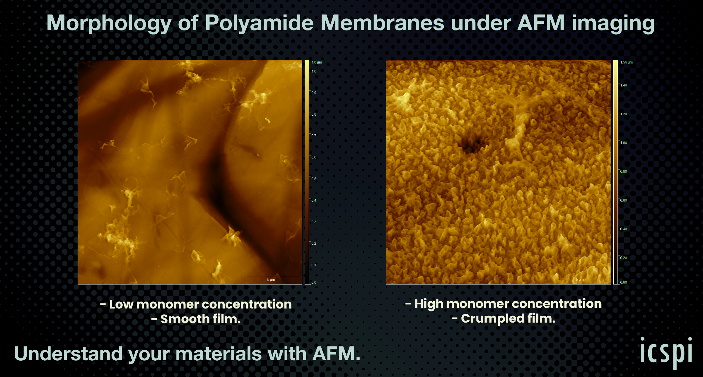
AFM for Reverse Osmosis Membranes
Atomic force microscopy for polyamide nanofilms in reverse osmosis membranes
What’s the Secret to Better Reverse Osmosis (RO) Membrane Performance?
It all starts with understanding the nano and microscale structure of the polyamide (PA) layer—the key to unlocking energy-efficient water separation.
This is what Professor Jay Werber and his PhD student Niher Ranjan Sarker from the University of Toronto were curious to better understand using atomic force microscopy (AFM). They fabricated two different polyamide nanofilm samples — both made from the same chemistry but with different monomer concentrations — resulting in two very different nano/micro scale structures:
🔹 Crumpled Film: Higher monomer concentrations
🔹 Smooth Film: ~10x lower monomer concentrations
Using AFM, we were able to see significant differences in the topography of the end product resulting from changes in initial monomer concentration.

ICSPI's AFM systems for membranes characterization
On-Chip AFM provides unmatched access to the true 3D structure of polyamide RO membranes. For researchers and developers aiming to improve permeability, selectivity, or scaling resistance, understanding the real surface structure—not just the chemistry—is critical. AFM enables this understanding through direct measurement of surface roughness, topology, and other quantitative metrics across a wide range of conditions.
ICSPI’s AFM systems like the nGauge AFM and Redux AFM are ideal for quickly and accurately characterizing membrane samples, without the need for sample coating or operation in vacuum.
Want to read more?
To learn more regarding what critical details AFM can provide and how it compares to data from Scanning Electron Microscopy (SEM), please download our full 6-page app note.
Speak with an expert
Interested to learn more about how AFM can provide insight on your membrane samples?